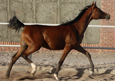
A useful decision begins before anyone tries to name the injury. A trainer may notice that a horse who used to turn smoothly now shortens a stride, takes longer to move forward, or sounds different on familiar ground. Such observations do not locate pain by themselves. They do, however, justify pausing before describing every resistance as disobedience or a fitness problem.
Lameness is a clinical sign, not a single disease. Its source may be the foot, a higher part of a limb, several limbs, or another system that alters movement. A short clip or comparison with another horse cannot establish a diagnosis. Veterinary guidance places history, physical examination and gait assessment first; the clinician then chooses localization tests and imaging when they are necessary and safe.
Urgency differs. A mild repeated change deserves an examination plan before hard work resumes. A sudden severe lameness, difficulty bearing weight, obvious swelling after an incident, or a suspected fracture calls for stopping movement and immediate veterinary contact. Forcing the horse to trot for a better video or finishing the session to confirm a suspicion can worsen a serious injury. Pain relief should not conceal the history; the veterinarian needs to know exactly what was given and when.
This guide gives the person responsible for the horse practical questions: what has changed from the animal's own baseline, and is it visible at walk, trot or under saddle? Is there heat, swelling, a wound or changed behaviour? What information should be saved for the veterinarian rather than tested at home? The goal is not to turn a rider into a lameness examiner. It is to reduce delay and supply an orderly history for an individual diagnostic and treatment plan.
1. Build a baseline for this horse
Observe the horse during familiar work while it is well, rather than collecting a baseline only after trouble begins. Record the approximate stride pattern, rhythm, transitions, ease of turning in both directions, standing posture and behaviour after rest. An expensive device is not essential: a dated note and a short recording on safe footing can support later comparison. Keep the surface, shoeing or barefoot status and date of the last farrier visit with the observation. These details shape interpretation without becoming diagnoses in their own right.
Compare the horse with itself, not with an abstract ideal gait. Some horses have stable conformational differences or familiar movement patterns known to their team. What matters is a new change, whether it repeats, and whether it is linked to a particular surface, direction or workload. If one person sees a problem, ask another for an independent description without suggesting a limb. That may reduce bias, but agreement between observers is not a substitute for examination. Disagreement is another reason to record conditions carefully for the veterinarian.
One trainer may dismiss an uneven step as fatigue; another may interpret every normal variation as an injury. Avoid both extremes with a written review rule. Has the pattern changed since last week, appeared on more than one pass, or coincided with declining performance or reluctance to do a familiar task? Do not increase the difficulty of exercise to test the answer. If uncertainty remains after safe observation, speak to the veterinarian before the next demanding session. The threshold is a decision to seek assessment, not a home-made diagnosis.
Use recording without treating video as a verdict. If the horse is already moving safely, film the whole body from the side and, when practical, from the front or rear. Record the footing, date, gait, handler and whether the clip was before or after warm-up. Do not force a tight circle or slippery surface to make an asymmetry more visible. Do not repeat passes once pain appears severe. The veterinarian decides whether more movement is required, and under what conditions. A useful clip preserves context rather than proving a theory.
2. Read the signals without guessing the cause
A forelimb problem can appear as a repeated head movement, while pelvic movement may change with hindlimb lameness. These are observational patterns used by clinicians within a broader examination, not a way to name the injured limb from a few seconds of footage. A horse may redistribute weight because several areas hurt. A skilled rider can also smooth an uneven rhythm without intending to conceal it. Describe what you actually see: a head movement, shorter step or broken transition. Leave localization to the veterinarian.
Look beyond rhythm for fresh swelling, local heat, a wound, a shifted shoe, reluctance to lift a foot or a changed weight-bearing posture. Compare sides gently and consistently, but do not press hard on a painful area or lift a foot when the horse cannot bear weight on the other one. Heat or a stronger digital pulse may direct the clinician's attention towards the foot; it does not prove an abscess or rule out laminitis or another injury. Record the observation. Do not cut into the hoof or remove a shoe on your own.
Behaviour matters when it is new and specific. A horse may hesitate over a familiar movement, change its balance under saddle, resist a turn or perform less consistently. Not every behaviour is caused by pain; there are other possibilities. Yet punishing the horse or demanding a stronger effort before checking health, tack and working conditions is a poor test. Veterinary literature treats subtle performance complaints as legitimate reasons for assessment, especially when an uneven gait is difficult to see from the ground. Record the task, direction and first date of change.
Pay attention to absences as well. No visible swelling does not prove that tissues are sound, and a straight-line walk that looks even does not show how the horse feels in a turn or under a rider. Conversely, a different hoof sound or a single stumble does not establish an injury. A useful picture combines the date of onset, recurrence, footing, workload, physical signs and hoof condition. Each piece improves the question put to the clinician. None licenses an experimental treatment or a strenuous self-test while the underlying issue remains unknown.

3. Know when movement must stop
If lameness starts suddenly and the horse can barely bear weight, or follows a fall or collision with swelling or deformity, safety comes first. Stop work, keep the horse in a secure place that does not require unnecessary movement, and contact the veterinarian immediately. Do not ask for a trot or a tight turn to grade the lameness. Veterinary guidance warns that exercising a horse with a possible fracture can cause catastrophic failure. The clinician should direct examination, stabilization and any transport plan.
Painful heat in the feet with a changed stance or obvious difficulty moving merits urgent contact. Laminitis is one possibility that should not wait for another working day, though it is not the only diagnosis. Nor should an acute lameness be declared 'just an abscess' because abscesses are common. Early signs can overlap between conditions requiring very different responses. A photograph of the hoof is not a treatment plan. Do not make the horse walk to ease what you assume is stiffness; obtain individual instructions from the veterinarian.
Even when the change seems milder, stop the session if it clearly repeats or worsens with work. Repeated loading to let the horse 'work out of it' can obscure or aggravate a developing problem. Enter the event in the training record, alert other handlers so no one resumes hard work unknowingly, and arrange veterinary assessment before returning to high load. If the sign disappears after minutes, keep the record: describe when it began and when it faded. That fluctuation may help the clinician, but it does not establish that continued work is safe.
While waiting, do not start painkillers, anti-inflammatory drugs or supplements to restore performance without veterinary direction. Medication can alter the clinical picture, and its timing matters when examination findings are interpreted. If a drug has already been given, record its name, amount, time and reason and tell the veterinarian openly. Do not hide the information for fear of criticism. Equally, do not impose a long period of strict confinement or a generic therapeutic exercise routine on your own. Management differs between acute and chronic conditions and between hoof, tendon and joint problems.
4. Prepare a useful history, not home tests
Prepare a short chronology beginning with the last day the horse moved normally. Include the first observed change, the work that preceded it, footing, any fall or slip, and whether walk, trot or ridden work showed a difference. Add recent trimming, shoeing or a shifted shoe and any relevant change in yard or routine. The veterinarian benefits more from verifiable timing and observations than from a long list of diagnostic possibilities assembled online. Keep the account factual even when the team disagrees about which leg appears affected.
Look from a comfortable distance, then touch limbs gently only if the horse is calm and can tolerate handling. Photograph a swelling or wound with a suitable size reference, and use a similar position and light for later comparison. Avoid forceful pressure and tests that cause pain or put the handler at risk of a kick. Flexion tests, hoof testers and diagnostic local anaesthesia are clinical tools selected and interpreted by professionals. Their results cannot be read apart from the whole examination and should never be improvised in the stable to prove a theory.
Give the clinician the work and injury history, medication record, farrier dates and earlier recordings, then ask what further footage is needed. Depending on severity, the veterinarian may watch a straight line, a circle or ridden movement; a suspected fracture changes what movement is safe. The examination may include foot, back and neck before localization and imaging are considered. The first radiograph does not necessarily settle every problem. A useful test answers a clinical question developed from history and examination rather than a wish to scan everything at once.
Do not be surprised if the veterinarian revises a working hypothesis as the examination progresses. Pain in one limb can produce compensatory changes elsewhere, and a ridden asymmetry does not locate the lesion alone. Ask which findings are established, which remain possibilities, why the next step is proposed and how response will be reviewed. Those questions are more useful than demanding a disease name after the first glance. Keep the visit record and safety instructions in the horse's file and share one consistent version with every handler so daily management does not fragment.

5. Return to work against documented criteria
Elapsed days alone should not be the return-to-work criterion. The veterinarian identifies the condition and stage of recovery, explains what activity is allowed and states which signs require stopping or reassessment. A horse recovering from soft-tissue injury may need a different approach from a horse with a foot or joint problem. Do not copy a rest period or walking programme from another case. Put each stage and its transition criterion in a record understood by trainer, rider and daily carer, and set a review date that is not merely the date of the next competition.
During return, compare movement, comfort and willingness to work with the recorded baseline rather than one good day. Note what happens after an increase in load and on the following day, because some complaints emerge later. Give the team a simple reporting format: date, work, surface, observed change and a recording where useful. If unevenness returns or swelling or behaviour changes, pause the progression and consult the veterinarian. Do not use pain relief so the horse can pass a functional test; that distorts the recovery measure and puts welfare second.
After diagnosis, review modifiable loads: session density, recovery days, footing, warm-up, farrier schedule and tack fit. Changing one factor is not assumed to cure the underlying condition; this is risk management and observation, not a replacement for treatment. Make each change traceable: what was altered, when, and whether movement improved without discomfort increasing. Do not move directly from a long rest to the previous workload because a few steps down the aisle looked even. The sporting decision must be aligned with the veterinarian's findings and the horse's actual response.
Judge the plan by outcomes meaningful for the horse: comfortable, repeatable movement, the ability to do the required task without new resistance, and a stable response to agreed load increases. The absence of visible lameness in one clip is not proof that an injured structure has healed. Keep the record when trainer or owner changes; otherwise the case loses its context and experiments begin again. This framework cannot promise that every injury will be prevented. It makes return to sport a reviewable decision rather than a hidden gamble under pressure from a race or show date.
6. Build a culture of reporting, not concealment
The person grooming or leading the horse may notice a change before anyone reviewing training figures does. Make reporting easy and do not penalize a good-faith concern; ask for a description and date, not a diagnosis. When a rider says that a horse no longer feels comfortable in a particular transition, do not translate the report immediately into disobedience. Ask what changed in direction, surface, tack and warm-up, then retain the question for professional assessment if it remains unexplained. Early reporting is useful precisely because it occurs before certainty is possible.
Use one incident record rather than scattered messages. At first, it needs the horse's identity, date, activity, footing, specific observation, severity described without an invented score, the decision to stop work, who contacted the veterinarian and what advice was given. Separate 'I saw swelling' from 'I think this is a tendon injury': one is an observation, the other a hypothesis. That distinction protects the clinical assessment from premature anchoring. It also helps the team see whether a sign is genuinely new or part of a pattern already recorded.
Do not turn monitoring into a contest over numbers. A sensor or slow-motion clip may highlight a change, yet measurements depend on pace, footing, operator and camera setup and require clinical interpretation. If data shift while the horse appears comfortable, review recording quality and the baseline with the veterinarian rather than changing training automatically. If movement or behaviour remains concerning despite a reassuring number, take the clinical observation seriously. A tool supports communication and comparison; it is not a permission slip to finish a session when the horse is giving a warning.
Review each case once it has been assessed. Did the team notice the sign early? Did finishing a session cause delay? Was the veterinary contact number available? Did records distinguish what was examined from what remained unknown? A review may lead to a modest change in shoeing notes or recovery-day planning, or no change if evidence does not support one. The important outcome is that a legitimate concern is not concealed for fear of withdrawing from an event. Equine sports-medicine guidance places welfare first; that principle is tested by small decisions made on the training ground.
Aletaq Golden AdviceIf the horse's rhythm changes from its normal pattern and the change repeats, stop exertion, record the circumstances and call the veterinarian. If pain is severe, weight-bearing is difficult or fracture is possible, do not test at trot: seek urgent help.
A decision supported by evidence
Early lameness calls for disciplined observation, not bold guessing. A stride change that repeats under the same conditions may be enough to end today's session while its cause remains an open question for the veterinarian. Record the horse's normal pattern when it is comfortable, then describe the new change in comparable terms: onset, footing, work and associated signs. Do not let an approaching event become a reason to increase load as a test or to mask pain with medication.
In a severe case or when weight-bearing is difficult, safety and urgent veterinary contact come first. With a subtle change, pausing, documenting and arranging an early examination turn uncertainty into a responsible decision. On return, ask for functional criteria specific to this horse rather than a number of days copied from another. What small sign in your own horse's movement do you wish you had recorded sooner? Add the answer to its health file, and subscribe to Aletaq's newsletter for carefully sourced practical guides when they appear.
Subscribe to Aletaq’s newsletter